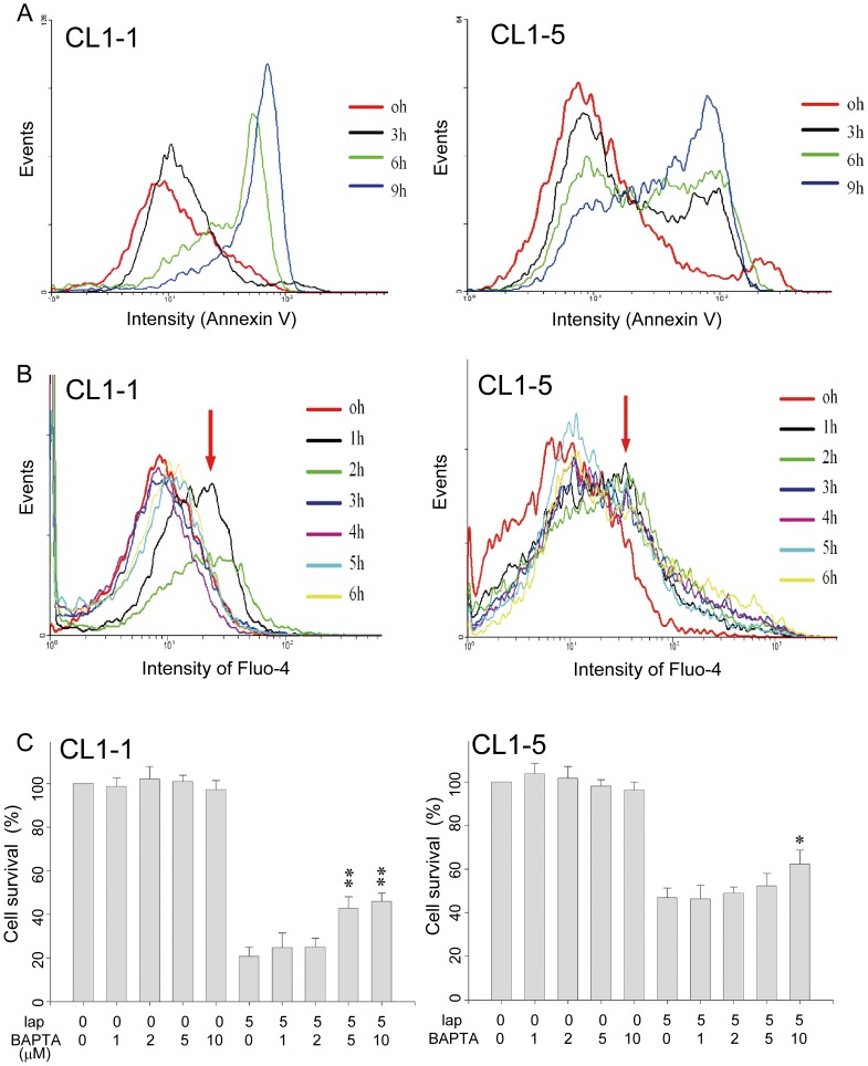
Figure 2. The β-lapachone-induced apoptosis of CL1-1 and CL1-5 cells is partly due to an intracellular calcium increase.
(A) CL1-1 cells (left panel) or CL1-5 cells (right panel) were incubated with 5 µM β-lapachone for 0, 3, 6, or 9 h, then were examined for apoptosis using Annexin V. (B) CL1-1 cells (left panel) or CL1-5 cells (right panel) were incubated with 5 µM β-lapachone for the indicated time, then intracellular calcium levels were measured using Fluo-4 staining and flow cytometry. The intensity of Fluo-4 staining was increased by β-lapachone treatment, especially at 1 h (arrows). (C) CL1-1 cells (left panel) or CL1-5 cells (right panel) were left untreated or were incubated for 24 h with the indicated concentration of BAPTA-AM, an intracellular calcium chelator, and/or 5 µM β-lapachone, then cell survival was measured by the MTT assay and expressed as percentage survival compared to the untreated cells. * p<0.05, ** p<0.01 as compared to β-lapachone alone.

