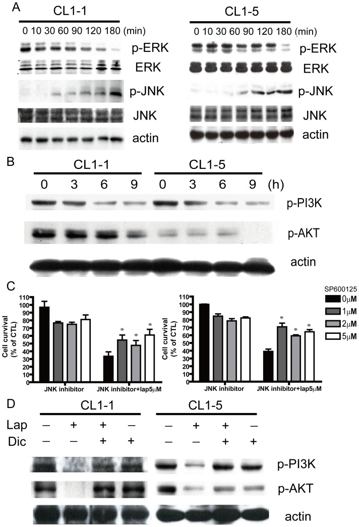
Figure 3. Signaling pathway components involved in β-lapachone-induced apoptosis.
(A) CL1-1 cells (left) or CL1-5 cells (right) were incubated with 5 µM β-lapachone for the indicated time, then levels of p-ERK, ERK, p-JNK, and JNK were measured by Western blotting. (B) CL1-1 cells (left) or CL1-5 cells (right) were incubated with 5 µM β-lapachone for 0, 3, 6, or 9 h, then levels of p-PI3K and p-AKT were examined by Western blotting. (C) CL1-1 cells (left) or CL1-5 cells (right) were pretreated with the indicated concentrations of the JNK inhibitor sp600125 for 6 h, and then treated with or without 5 µM β-lapachone for 24 h.Cell survival was measured by the MTT assay and expressed as percentage survival compared to the untreated cells.* p<0.05 (D) CL1-1 cells (left) or CL1-5 cells (right) were left untreated or were preincubated for 1 h with 10 µM dicoumarol, then medium or 5 µM β-lapachone was added and the cells incubated for 9 h and levels of p-PI3K and p-AKT were measured by Western blotting.

