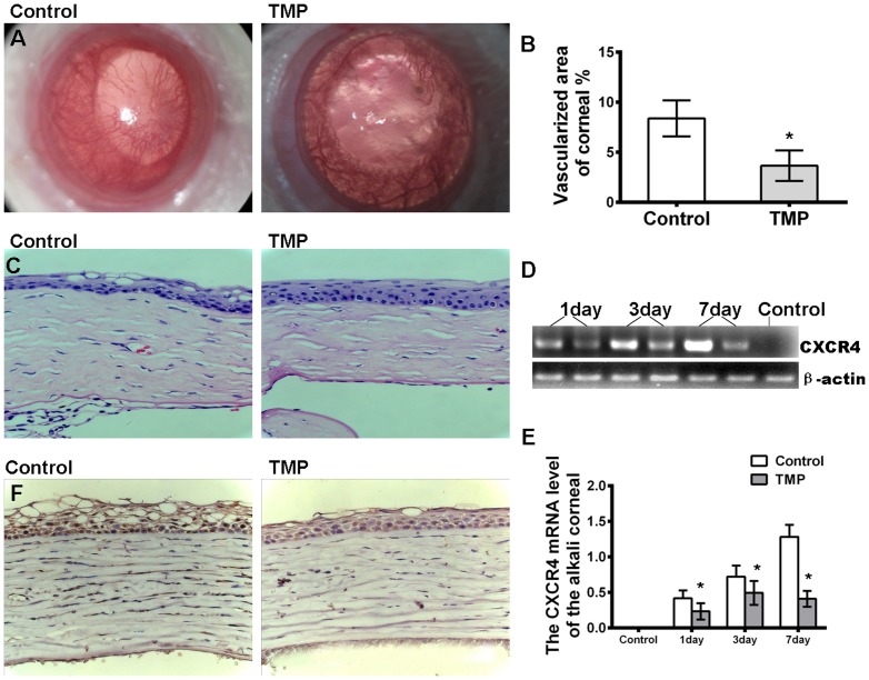
Figure 2. Effects of TMP on corneal NV.
(A) Alkali-induced corneal neovascularization is suppressed through TMP treatment. Photos were taken at day 28. (B) The bar graph represents the mean and SE of the blood vessel within the microscopic fields from 15 eyes. (C) The hematoxylin and eosin staining of alkali-burned corneas at 28 days post-injury shows that stromal inflammation and edema (evaluated by the stromal thickness) are reduced in the corneas of the animals in the TMP-treated group compared with those in the control group. (D) The RT-PCR analysis of CXCR4 mRNA in the cornea post-injury. These results are representative of experiments performed at least three times. (E) The relative level of CXCR4 mRNA was quantified through densitometry. (F) Immunohistochemical staining indicates that CXCR4 (brown) is reduced in the cornea at 7 days after TMP treatment. The asterisks represent statistically significant differences between TMP and the control (*p<0.001).

