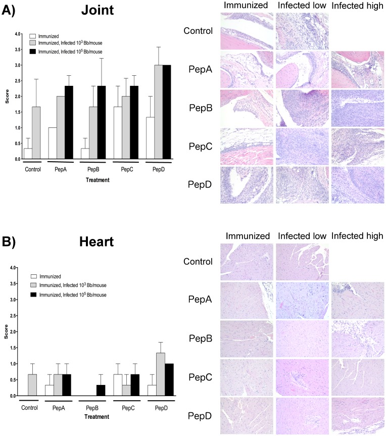
Figure 2. Representative histological images of the average level of inflammation observed in each treatment group (control, pepA, pepB, pepC, and pepD) after immunization and/or infection with the low (103 spirochetes/mouse) or high (105 spirochetes/mouse) doses in the tibiotarsal joint (A) and the heart (B).
Tissues were histologically evaluated at four weeks post priming, as well as four weeks post needle inoculation. Average scores for areas of inflammation were classified as 0 = none; 1 = minimal; 2 = mild; 3 = moderate; 4 = severe. Peptide B induces minimal inflammation in hearts and tibiotarsal joints after administration in the mouse model for Lyme disease. Of all the peptides evaluated after immunization, only peptide B showed inflammation comparable to the negative control group in both heart and joints. Similar results were observed after infection with low doses of B. burgdorferi. Images were captured using an Olympus BX41 microscope at 200X magnification. Average ± SD are presented in the graphs.

