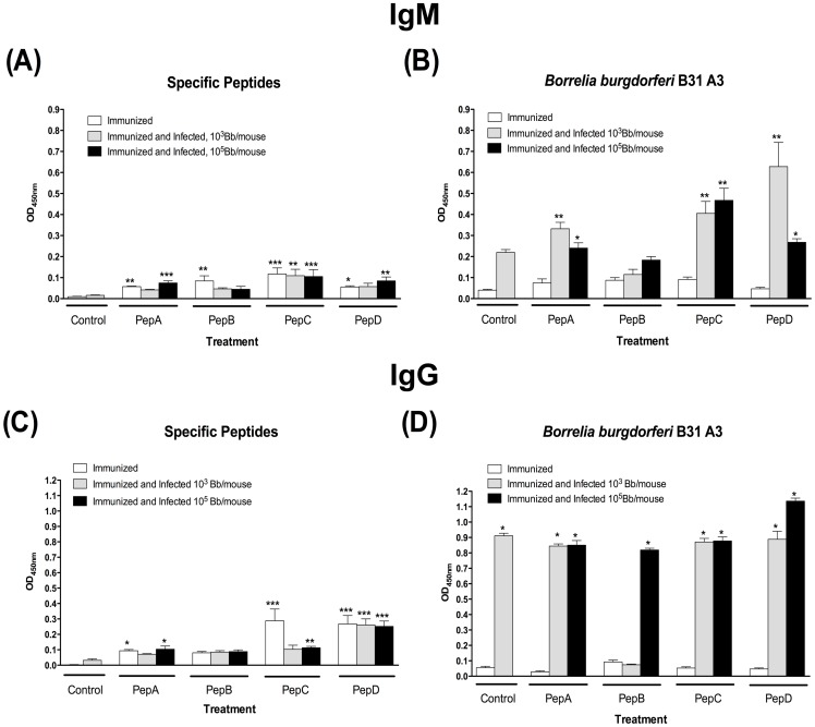
Figure 3. Low IgM and IgG antibodies were detected 4-weeks post priming in all groups.
Antibody levels were evaluated 4-weeks post-priming as well as 4-weeks post needle infection. (A) Peptide-specific IgM antibodies. (B) B. burgdorferi-specific IgM antibodies. (C) Peptide-specific IgG antibodies. (D) B. burgdorferi-specific IgG antibodies. * Denotes statistically significant differences (* P value <0.05; ** P value < 0.01; *** P value < 0.001) when compared with the control group.

