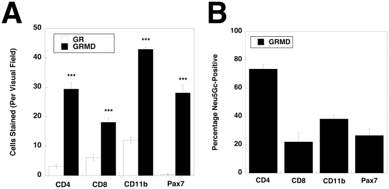
Figure 5. Quantification of CD4−, CD8−, CD11b− and Pax7−stained cells in GR and GRDM muscle and of Neu5Gc co-staining in GRMD muscle.
(A) Cranial sartorius muscle sections from GR and GRMD dogs were stained with markers for satellite cells (Pax7), T lymphocytes (CD4 or CD8) or macrophages (CD11b) and quantified for numbers of cells stained per 40X visual field. (B) The percentage of cells co-stained for Neu5Gc and CD4, CD8, CD11b or Pax7 in GRMD muscles was quantified. Errors are SEM. ***P<0.001, for each GR vs. GRMD comparison in A.

