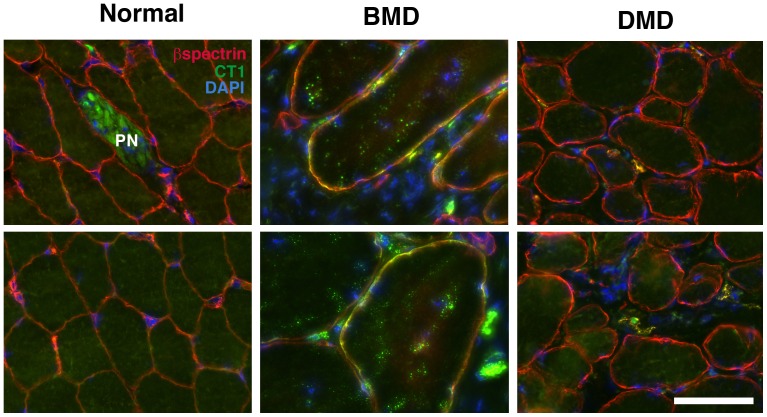
Figure 10. Co-staining of CT1 with βspectrin in BMD muscles.
Sections from DMD, BMD and normal human muscle biopsies sections were co-stained with CT1 (green), β spectrin(red) and DAPI (blue). Merged sarcolemmal membrane staining for CT1 and β spectrin is orange-yellow. PN represents intramuscular peripheral nerve. Bar is 50 µm for all panels.

