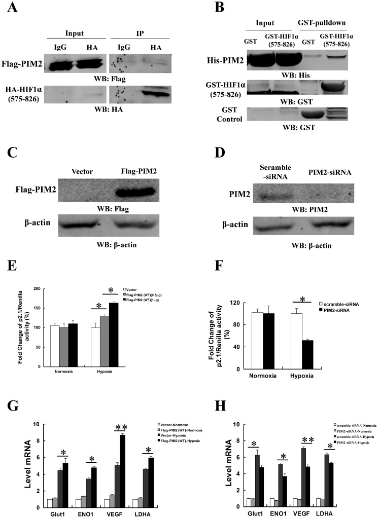
Figure 4. PIM2 augments HIF-1α–mediated transcription under hypoxia.
(A) HEK293T cells were transfected Flag-tagged PIM2 with HA-tagged HIF-1α (575-826aa) for 24 h and cultured under hypoxia for a further 24 h. Co-IP assays were performed with anti-HA antibody, followed by immunoblot assays. (B) GST pull-down assays were performed with GST, GST-tagged HIF-1α (575-826aa) and His-tagged PIM2 proteins which were purified from Ecoli expressing system. (C and D) HepG2 cells were transfected empty vector or Flag-tagged PIM2 (C); scramble siRNA or PIM2 siRNA (D) for 24 h and cultured under normoxia or hypoxia for a further 24 h. Protein Levels of PIM2 were determined in immunoblot assays using the indicated antibodies. (E and F) HepG2 cells were transfected empty vector or Flag-tagged PIM2 (E); scramble siRNA or PIM2 siRNA (F) with p2.1 luciferase reporter plasmid for 24 h and cultured under normoxia or hypoxia for a further 24 h before luciferase activity was measured. Transfection efficiency was normalized against Renilla luciferase expression. (G and H) HepG2 cells were transfected with empty vector or Flag-tagged PIM2 (G); scramble siRNA or PIM2 siRNA (H) for 24 h and cultured under normoxia or hypoxia for a further 24 h. mRNA levels of Glut1, ENO1, VEGF and LDHA were determined by real-time PCR assays. All data represent the means ± SEM of three independent experiments, *p<0.05, **p<0.01.

