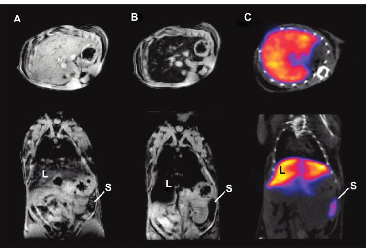
Figure 5.
(A–C) In vivo magnetic resonance (MR)–single-photon emitted tomography (SPECT) images of axial view (top) and coronal view (bottom).
Notes: (A) T2*-weighted MR images before injection of technetium-99m–diethylene triamine pentaacetic acid (99mTc-DTPA)-ale-Endorem; (B) T2*-weighted MR images 15 minutes after injection of 99mTc-DTPA-ale-Endorem; (C) SPECT image 45 minutes after injection of 99mTc-DPA-ale-Endorem; L indicates the liver, S the spleen, showing the accumulation of 99mTc-DPA-ale-Endorem at those organs with MR imaging and SPECT after injection. Reproduced with permission from Torres Martin de Rosales R, TavaréR, Glaria A, Varma G, Protti A, Blower PJ. (99m)Tc-bisphosphonate-iron oxide nanoparticle conjugates for dual-modality biomedical imaging. Bioconjug Chem. 2011;22(3):455–465.82 Copyright © 2011 American Chemical Society.

