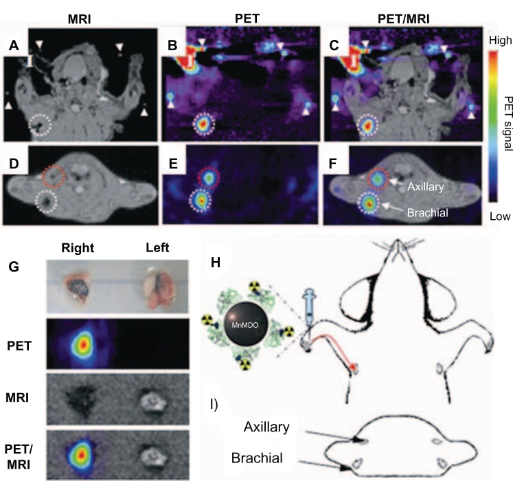
Figure 6.
(A–I) In vivo magnetic resonance (MR) and positron emission tomography (PET) images of sentinel lymph nodes in a rat 1 hour after injection of iodine-124–serum albumin–manganese magnetism-engineered iron oxide into the right forepaw.
Notes: Coronal images of (A) in vivo MR, (B) in vivo PET, (C) MR–PET fusion; transverse images of (D) in vivo MR, (E) in vivo PET, (F) MR–PET fusion; (H) explains the coronal images of (A–C); (I) describes transverse directions of (D–F); (G) ex vivo images of brachial lymph nodes by PET, MR imaging, and PET/MR imaging; (I) in (A–C) indicates the injection site. Reproduced with permission by Choi JS, Park JC, Nah H, et al. A hybrid nanoparticle probe for dual-modality positron emission tomography and magnetic resonance imaging. Angew Chem Int Ed Engl. 2008;47(33):6259–6262.

