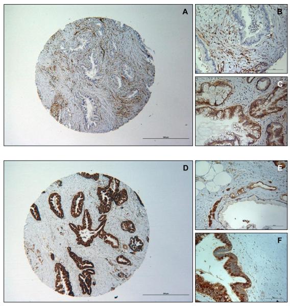
Figure 2. Loss of PTEN expression and overexpression of PDK1 in IPMN.
(A) Representative absent immunopositivity of PTEN (40X); (B) Example of absent PTEN expression in IPMN with strong immunolabeling in stroma (200X). (C) Example of normal/strong cytoplasmic expression of PTEN in IPMN (200X). (D) Representative strong immunopositivity of PDK1 (40X). (E) Detail of weak cytoplasmic PDK1 immunolabeling in IPMN (200X). (F) Example of strong cytoplasmic PDK1 immunopositivity in IPMN (200X). Scale bars are 500um and 100um.

