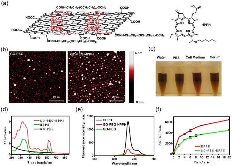
Figure 1.
a) Schematic structure of GO-PEG-HHPH. b) AFM images of GO-PEG and GO-PEG-HPPH dispersed in ultra-pure water. Scale bar: 250 nm. c) Stability of GO-PEG-HPPH in water, PBS, cell medium and serum. No precipitation was observed at 24 h after incubation. d) Normalized UV-vis spectra of GO-PEG, HPPH and GO-PEG-HPPH. Two characteristic absorption peaks at 414 and 665 nm were observed for GO-PEG-HPPH. e) Fluorescence spectra of free HPPH, GO-PEG and GO-PEG-HPPH at a concentration of 1 µM of HPPH and 0.49 µg of GO-PEG. f) Singlet oxygen generation of free HPPH and GO-PEG-HPPH (1 µM) after irradiation with 671 nm laser (75 mW/cm2) for different periods of time.

