Figure 3.
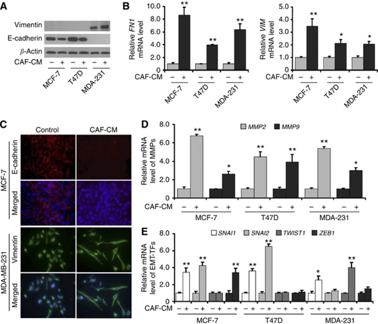
CAF-CM induces EMT programming and phenotype in different breast cancer cell lines. (A) The expression of epithelial marker E-cadherin and mesenchymal marker vimentin was detected by immunoblotting. Compared with control cells, the cells cultured with CAF-CM had decreased E-cadherin expression in MCF-7 and T47D cells, and increased vimentin expression in MDA-MB-231 cells. (B) The expression of mesenchymal marker VIM and FN1 was detected by reverse transcription (RT)-quantitative PCR (QPCR). Compared with control cells, VIM and FN1 expression levels were upregulated in all the three cell lines cultured with CAF-CM. (C) The expression of epithelial marker E-cadherin and mesenchymal marker vimentin was detected by immunofluorescence staining. Compared with control cells, the cells cultured with CAF-CM had decreased E-cadherin expression in MCF-7 cells and increased vimentin expression in MDA-MB-231 cells cultured with CAF-CM. Nuclei were visualised with DAPI staining (blue). (D) The expression of MMPs was detected by RT-QPCR. MMP2 and MMP9 expression levels were upregulated in all the three cell lines cultured with CAF-CM. (E) The expression of EMT-TFs was detected by RT-QPCR. SNAI1 expression levels were upregulated in all the three cancer cell lines cultured with CAF-CM. SNAI2 expression levels were upregulated in MCF-7 and T47D cells. TWIST1 expression levels were upregulated in MDA-MB-231 cells, and ZEB1 expression levels were upregulated in MCF-7 cells. *P<0.05, **P<0.01.

