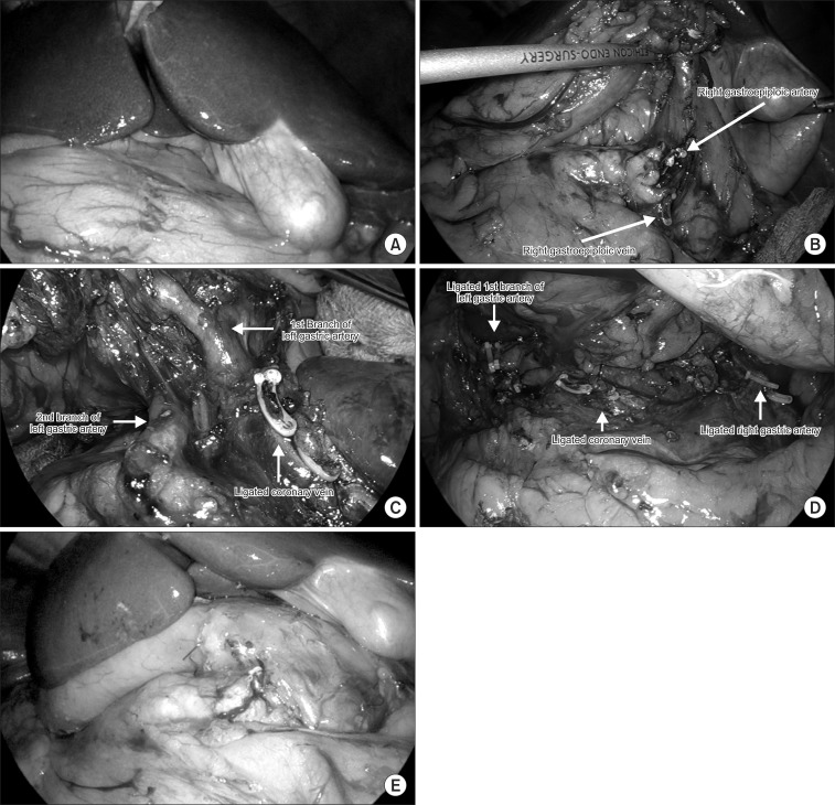
Fig. 4.
Case 1. (A) Initial laparoscopic view showing transposition of abdominal organs. (B) The ligated right gastroepiploic artery and vein. The ligated coronary vein. (C) Anatomic variation in the 1st and 2nd branch of the left gastric artery is apparent. (D) The ligated 1st branch of the left gastric artery, right gastric artery and coronary vein. (E) The wound after Billroth I anastomosis and Fibrin glue had been applied.

