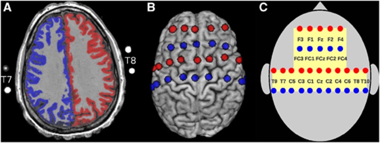
Figure 3.
(A) Representation of the vascular territories supplied by the left (blue) and right internal carotid artery (ICA) (red) as assessed by selective arterial spin labeling (ASL) on the anatomy of a healthy subject. A cortex mask was applied to restrict the representation to cortical structures. Outside the skull magnetic resonance (MR)-visible markers (nitroglycerin medication capsules) of some near infrared spectroscopy (NIRS) optode positions are visible. (B) Projection of the optode positions (transmitters in red and receivers in blue) onto the skull-stripped brain of the same subject. Lateral views are available in Supplementary Figure S1 of the Supplementary Information. (C) Schematic representation of the optode montage with reference to approximate positions in the 10/10 electroencephalography system. Each optode pair spaced at 30 mm constitutes an NIRS channel. The yellow-shaded region is monitored by NIRS. This spatial layout is used in Figure 6 for representation of the results in all patients.

