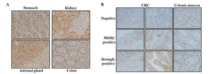
Figure 2.

Immunohistochemical (IHC) analysis for heparan sulfate 6-O-sulfotransferase-2 (HS6ST2) in (A) normal tissues and (B) representative images of colorectal cancer (CRC) tissues. IHC stainings of the stomach, adrenal gland and kidney are shown, all of which are representative examples of a strong expression. No expression was observed in normal colonic tissues. The results of the normal tissue array are summarized in Table I. Representative IHC staining in colonic tissues of CRC patients is defined as negative, mildly positive and strongly positive, according to the expression level and staining intensity.
