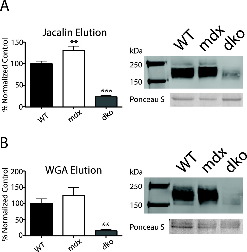
Figure 3. Lectin binding assays.
Mouse cardiac homogenate proteins bound to jacalin- (a) or WGA- (b) conjugated agarose beads. Proteins were eluted, subjected to SDS-PAGE, and probed for α-DG on a western blot. α-DG expression is reported as a normalized percentage of WT α-DG signal +/− SEM. Significantly less dko α-DG was eluted from both WGA and jacalin lectins compared to WT, whereas mdx α-DG eluted more α-DG from the jacalin lectin compared to WT α-DG. Ponceau S stained band (~70 kDa) provided as a loading control. (n = 4–9) ** p < 0.001, *** p < 0.0001

