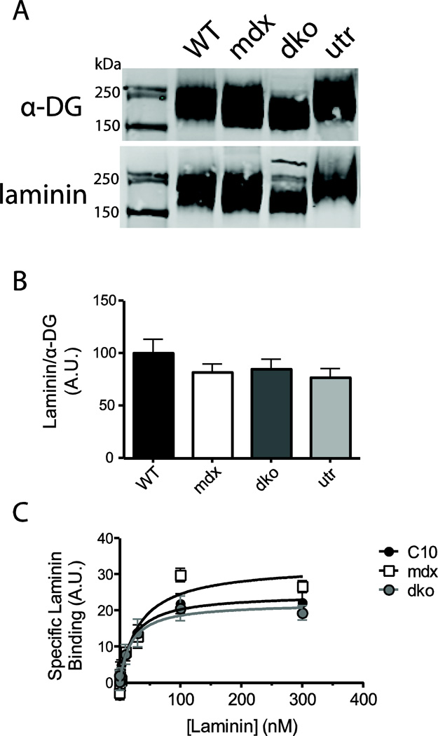
Figure 4. Laminin overlay binding assay.
The laminin binding capacity of α-DG was assessed using a far-western technique (a), and results were quantified by calculating the raw laminin signal over the raw α-DG signal (b). A solid-state binding assay was used to determine the affinity the laminin binding sites in cardiac homogenates (c). No significant difference in laminin binding capacity or affinity was observed between WT, mdx, dko and utr α-DG. (n=7–8)

