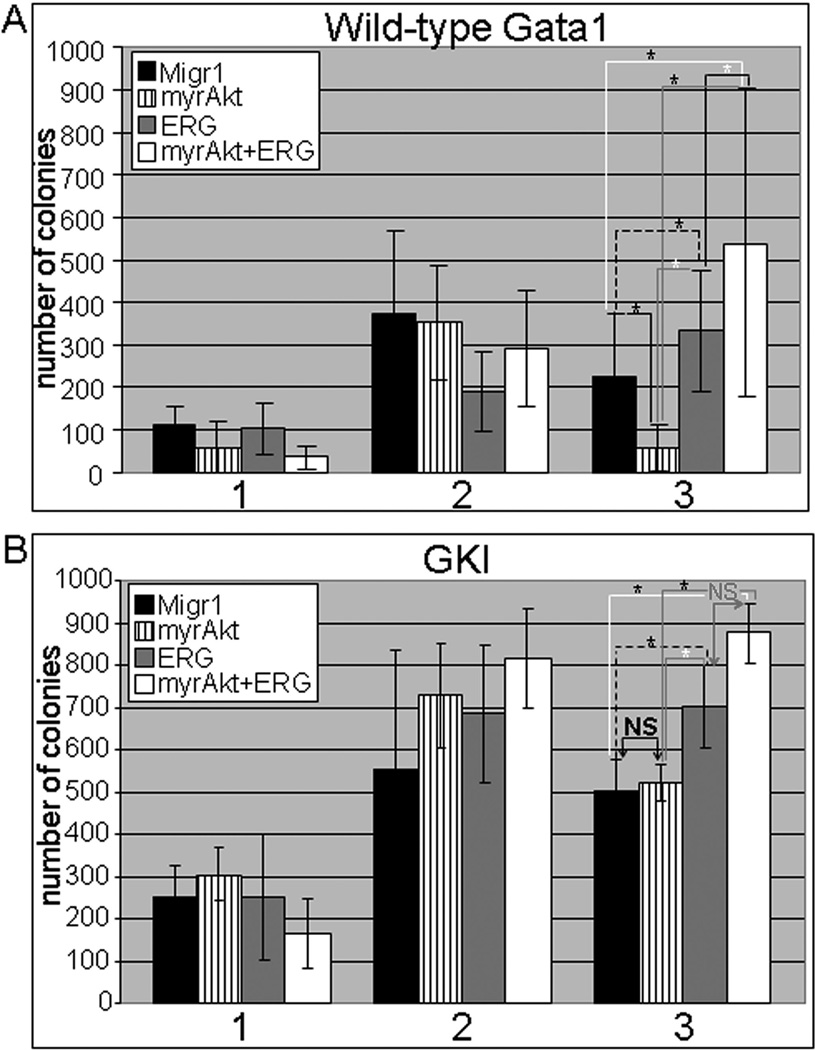
Figure 2. Long-term culturing demonstrates enhanced survival in the presence of ERG and GATA1s for myrAKT expressing cells.
(A) Serial replating of 5000 sorted GFP, mcherry double positive wild-type GATA1 (top) and (B) Gata1s knock-in (GKI) fetal liver hematopoietic progenitors (bottom); For wild-type GATA1 samples, p-value ≤ 0.04 for all * shown in figure. For GKI, p-value ≤ 0.04 for all * shown in figure. NS=not significant. The Student’s t-test was used for determination of statistical significance between each pair of linked samples. N=4 independent experiments for the wild-type cells and N=3 for the G1KD progenitors.

