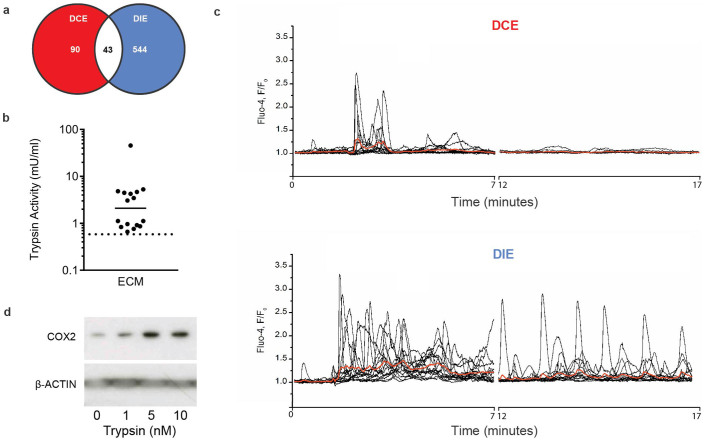
Figure 3. Competent pre-implantation human embryos actively induce a supportive uterine environment.
(a) Venn diagram presenting the number of maternal genes significantly (P < 0.01) altered 24 h after exposure of mouse uterus to unconditioned embryo culture medium or medium conditioned by either developmentally competent or impaired human embryos (DCE and DIE, respectively). (b) Tryptic activity was measured in 16 cultures containing a total of 163 human embryos between day 4 to 6 of development. Activity in unconditioned medium was below dotted line. (c) Application of embryo-conditioned medium induces [Ca2+]i oscillations in Ishikawa cells. Black traces show [Ca2+]i recordings from individual cells in response to application of 1:20 diluted culture medium obtained from DCE (top panel) or DIE (bottom panel). Red traces represent average of all individual traces in each panel. (d) Total protein lysates obtained from Ishikawa cells 24 h after treatment with trypsin (10 nM) for 5 min were subjected to western blot analysis and immunoprobed for COX2. β-ACTIN served as a loading control. Full length images are presented as Supplementary Information.

