
Figure 1-4.
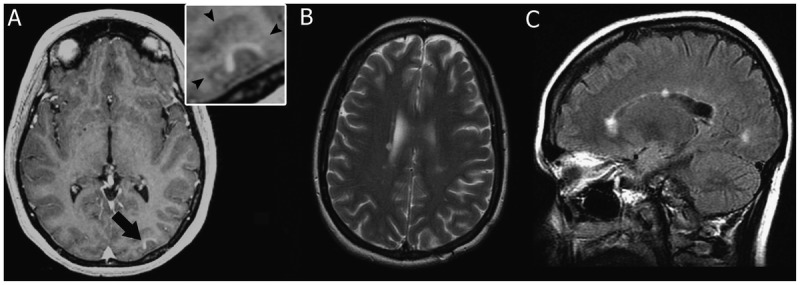
MRI of multiple sclerosis cortical onset. A, Prebiopsy axial T1-weighted image with contrast showing enhancement of the cortical lesion (arrow). Inset shows that contrast enhancement is within the cortical gray matter (arrowheads indicate the gray/white matter junction). B, Axial T2-weighted image 69 months after biopsy showing the appearance of new periventricular white matter lesions. C, Sagittal fluid-attenuated inversion recovery (FLAIR) image 69 months after biopsy showing the appearance of multiple new white matter lesions, with two of the lesions involving the corpus callosum.