
Figure 1-5.
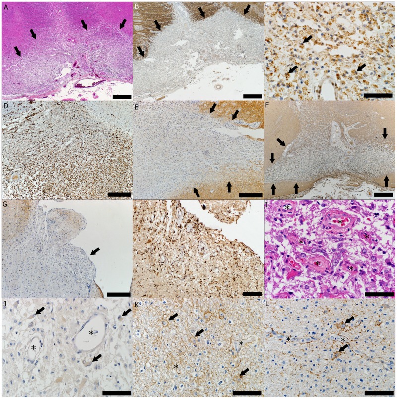
Pathology of a neuromyelitis optica (NMO) lesion located in the ventral medulla. A, Destructive hypercellular lesion; arrows indicate the border between the lesion that shows atypical hematoxylin and eosin (HE) staining (area below arrows) and normal-appearing white matter with preserved HE staining (area above arrows) (HE, scale bar = 500 μm). B, Demyelinated lesion; arrows indicate the border between lesion that shows loss of proteolipid protein (PLP) immunohistoreactivity (area below arrows) and normal-appearing white matter with preserved PLP immunohistoreactivity (area above arrows) (PLP immunohistochemical [IHC] staining, scale bar = 500 μm). C, Active demyelination (arrows indicate myelin-laden macrophages) (PLP, scale bar = 50 μm). D, The lesion shows macrophage infiltration (KiM1P IHC, scale bar = 250 μm). E, Loss of aquaporin-4 (AQP4) is characteristic for active NMO lesions; arrows indicate the border between lesion that shows loss of AQP4 immunohistoreactivity (area below arrows) and increased AQP4 immunohistoreactivity seen at the lesion’s edge (area above arrows) (AQP4 IHC, scale bar = 250 μm). F, Loss of glial fibrillary acidic protein (GFAP) is also frequently encountered in NMO-active demyelinating lesions; arrows indicate the border between the lesion that shows loss of GFAP immunohistoreactivity (area below arrows) and normal GFAP immunohistoreactivity of the normal-appearing white matter surrounding the lesion (area above arrows) (GFAP IHC, scale bar = 500 μm). G, Loss of aquaporin in the area postrema (arrow) (AQP4 IHC, scale bar = 250 μm). H, Macrophage/microglia infiltration in the area postrema (KiM1P IHC, scale bar = 100 μm). I, Area postrema blood vessels (asterisks) with thickened and hyalinized walls (HE; scale bar = 50 μm). J, Active NMO lesions with loss of AQP4 immunoreactivity seen as the disappearance of the perivascular rim or rosette AQP4 pattern (asterisks) and loss of AQP4 outlining of the astrocytic surface (arrows) (AQP4 IHC; scale bar = 50 μm). K, Increased AQP4 immunoreactivity outlining the cytoplasmic surface of reactive astrocytes (arrows) with preservation of the typical perivascular distribution (asterisks) at the border of the NMO lesion (AQP4 IHC; scale bar = 50 μm). L, Increased AQP4 immunoreactivity outlining the cytoplasmic surface of reactive astrocytes (arrows) with preservation of the typical perivascular distribution (asterisks) within an active multiple sclerosis lesion (AQP4 IHC; scale bar = 50 μm).