Fig. 4.

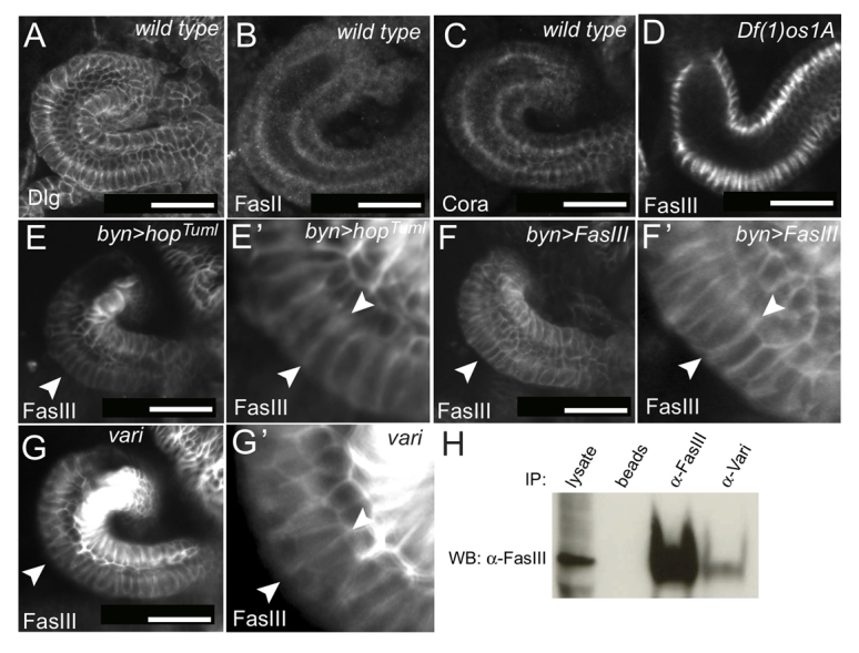
FasIII subcellular localisation. Dorsal views of stage 13/14 hindguts of the indicated genotypes and stained with the indicated antibodies. (A-C) Septate junction proteins Discs Large (Dlg), Fasciclin II (FasII) and Coracle (Cora) are symmetrically distributed and localised in sub-apical regions. (D-E′) Overview and magnification of FasIII localisation in stage 13 hindguts in JAK/STAT loss- and gain-of-function backgrounds. Arrowheads indicate examples of lateralised FasIII. (F,F′) FasIII is lateralised around the outside of a stage 13 hindgut (arrowheads) following its overexpression. (G,G′) Asymmetrical FasIII expression and ectopic lateralisation on the outside of the curve (arrowheads) in varicose (vari) mutants. (H) Co-immunoprecipitation from wild-type embryos using the indicated antibodies and blotted (WB) with anti-FasIII indicates a co-precipitation of FasIII with Vari. Scale bars: 30 μm.
