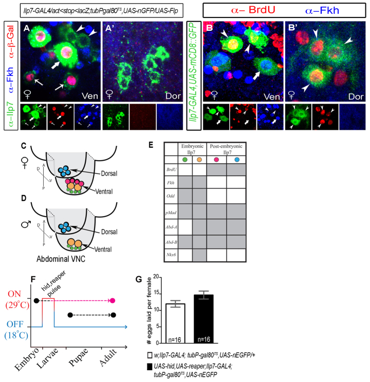
Fig. 2.
Post-embryonic Ilp7 neurons are sufficient for female fertility and can be distinguished from embryonic Ilp7 neurons by lack of Forkhead expression. (A-E) Expression of transcription factors in posterior Ilp7 neurons in adults. (A,A′) Transient Flp-induction-marked embryonic Ilp7 neurons with β-Gal. Forkhead (Fkh) was only expressed in embryonic Ilp7 neurons (arrows). (B,B′) BrdU incorporation into Ilp7 neurons in mid-late L3 larvae was only seen in Fkh-negative Ilp7 neurons (arrowheads) in ventral (Ven) and dorsal (Dor) clusters. Arrows/arrowheads indicate representative neurons of each Ilp7 subset. (C-E) Summary of transcription factor profile in embryonic and post-embryonic posterior Ilp7 neurons in adults (supporting data in supplementary material Fig. S2). Green circles indicate small embryonic Ilp7 neurons; yellow circles indicate large embryonic Ilp7 neurons; pink circles indicate the female-specific post-embryonic Ilp7 neurons; blue circles indicate the common post-embryonic Ilp7 neurons. (F,G) Selective killing of embryonic Ilp7 neurons does not disrupt female fertility. (F) Schematic of transient cell death gene expression in embryonic Ilp7 neurons (hid, reaper pulse). (G) Quantification of the number of eggs laid per female during a 6-hour assay following a 24-hour mating (mean±s.e.m.). Female fertility was not significantly different after killing embryonic Ilp7 neurons (black column), compared with control (white column) (control, 9.4±3.3; experimental, 11.8±5.7). n, number of egg-lay assays.

