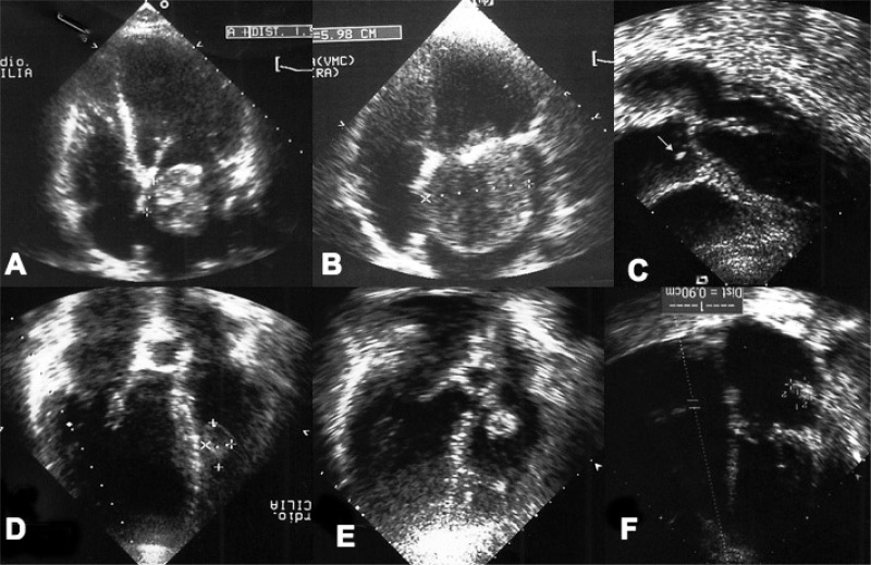
Fig. 1.
A, B) Preoperative echocardiography showing clearly the mechanism of obstruction of the mitral valve. C) Papillary fibroelastoma of the right aortic leaflet (white arrow) in a patient with previous embolic acute myocardial infarction. D) Papillary fibroelastoma of the right interventricular septum in a patient with malignant arrhythmia. E) Papillary fibroelastoma of the tricuspid valve. F) Recurrent myxoma of the left atrial wall after 29 months from the first surgery.

