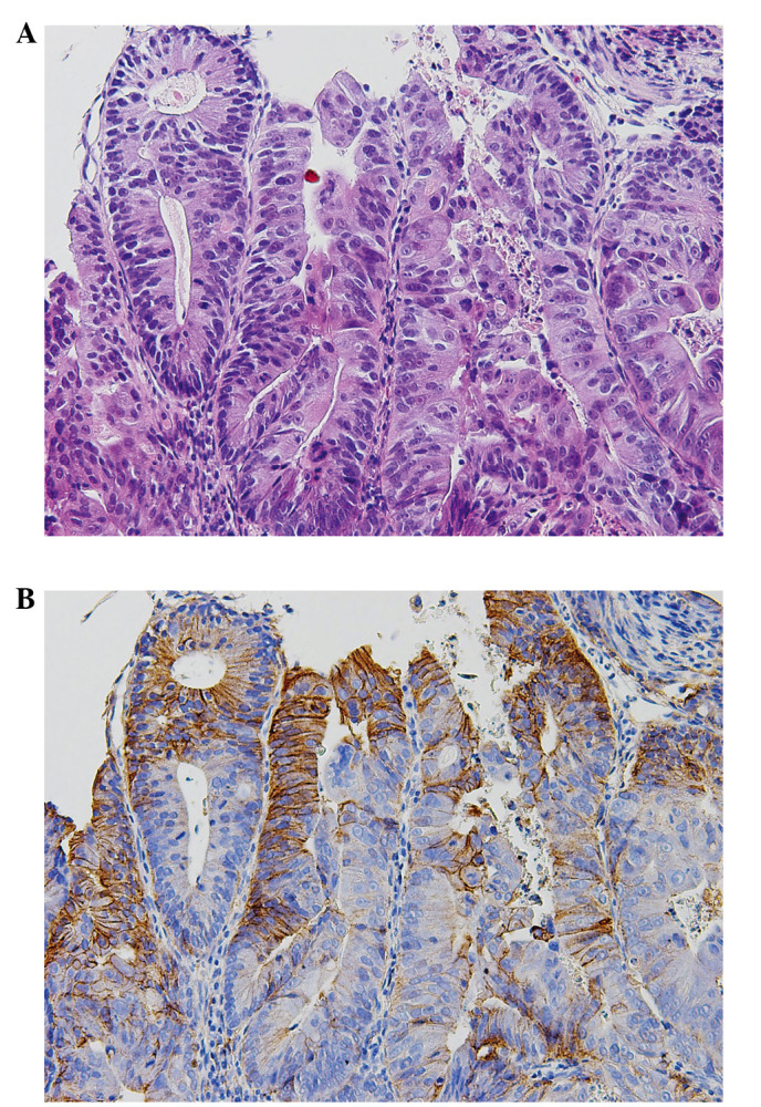
Figure 1.

Immunohistochemical staining of EGFR. (A) Hematoxylin and eosin shows a high-power view of moderately differentiated tubular adenocarcinoma of unresectable colorectal carcinoma. (B) Immunohistochemistry shows positive staining for EGFR (31G7) in the cytoplasmic membrane of carcinoma.
