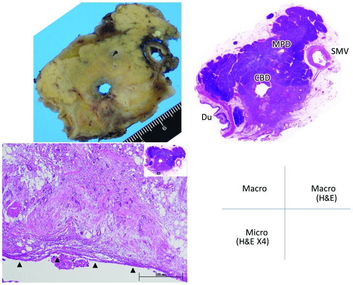
Figure 2.
Grade 1 retropancreatic infiltration by pancreatic head carcinoma. The specimen was cut perpendicular to the body axis. The tumor has infiltrated beyond the parenchyma and is adjacent to the fusion fascia. Arrowheads indicate the fusion fascia. Du, duodenum; CBD, common bile duct; MPD, main pancreatic duct; SMV, superior mesenteric vein. Macro, macroscopic view of the resected specimen; Macro H&E, macroscopic specimen stained with hematoxylin and eosin; Micro (H&E ×4), microscopic section stained with H&E, under magnification ×4.

