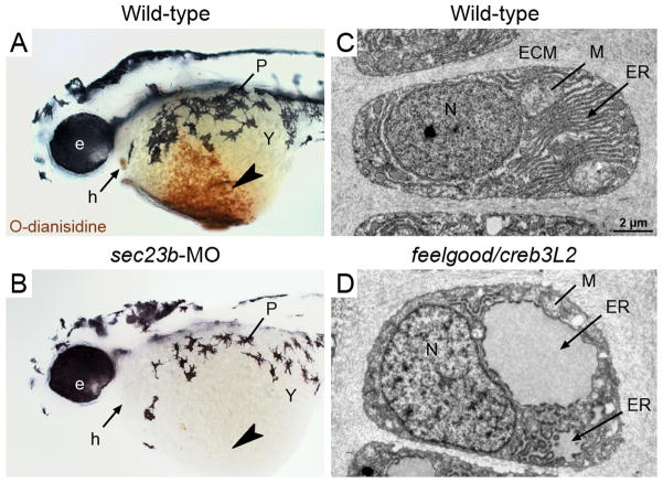
Figure 2. Deficits in secretory machinery lead to tissue-specific phenotypes.
A,B. Wild-type 2 day zebrafish embryos stained with o-dianisidine, which is oxidized by heme in the presence of peroxide and colored brown, display abundant hemoglobin within the ducts of Cuvier (arrowhead) and the heart (arrow). B. Sec23b morphants, which have defects in erythrocyte development, are deficient of hemoglobin at 2 days. C,D. Transmission Electron Micrographs (TEM) images of wild-type zebrafish chondrocytes (3-day old) show characteristics of highly secretory cells, including abundant rough ER (arrow), mitochondria (line), and Golgi. D. feelgood/creb3L2 mutant chondrocytes contain highly distended rER (arrow) due to collagen backlog. Symbols: e, eye; h, heart; P, pigment; Y, yolk; ECM, extracellular matrix; ER, endoplasmic reticulum; M, mitochondria; N, nucleus.

