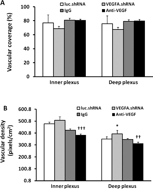
Figure 4.

Retinal vascular coverage and density in the inner and deep plexi of pups treated with VEGFA.shRNA or anti-VEGF in the ROP model at p18 compared with respective controls. (A) Retinal vascular coverage and (B) vascular density (*P < 0.05 versus luc.shRNA; ††P < 0.01 and †††P < 0.001 versus IgG, two-way ANOVA; results are means ± SD).
