
Figure 6.
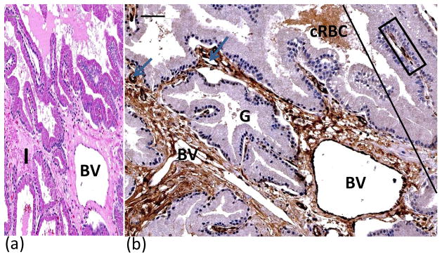
H&E (A) and vWF immunostained (B) sections illustrated effects of vascular damage in MZ. H&E staining showed loosely woven collagen and expansion of interstitial tissue due to leakage of plasma from damaged blood vessels resulting in edema(A). MZ is to the left and UZ to the right of tangential line (B). Blue arrows point to blood vessels in the interstitium. Note dark brown staining of endothelial cells lining dilated blood vessel (BV). Interstitium immunopositive for vWF due to leakage of plasma into interstitium from thermally injured blood vessels. Crenated red blood cells and plasma within lumen of prostatic gland were immunopositive for vWF. Immunopositive endothelial cells lining an intact vein (within blue rectangle) in UZ with no edema in interstitium. BV = blood vessel; cRBC = crenated red blood cells; G= prostatic gland. Bars (upper left) = 50μ.