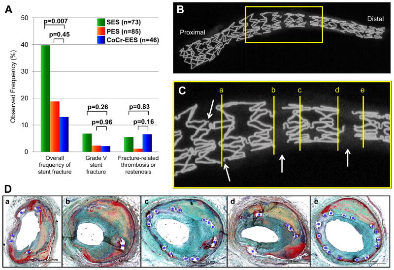
Figure 7.
Stent fracture in CoCr-EES. (A) Bar graph showing observed frequency of overall fracture, grade V fracture (acquired transection with gap in the stent body), and fracture-related thrombosis or restenosis, in CoCr-EES versus SES and PES. The analyses include duration of implant as a covariate. Multiple-comparison threshold is used as in Table 1. (B to D) A case of grade V CoCr-EES fracture showing restenosis at fracture site (Case #3 in Table 3). Radiograph of left obtuse marginal branch (B) shows a single CoCr-EES with multiple grade V fractures, which are highlighted in (C) (arrows=fracture sites). (D) Histologic sections of the fracture site (corresponding with [a] to [e] in panel C) showing focal restenosis. Note single stent struts (*) in the most severely narrowed section in b.

