
Figure 7. Electron Micrographs of N-MuSK 60-Immunized Animals.
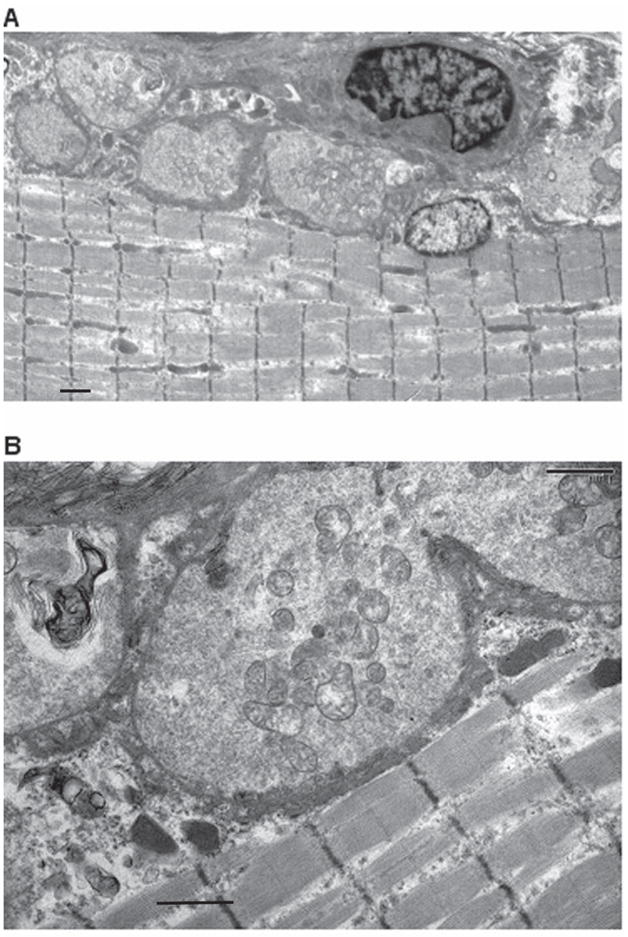
Electron micrographs of NMJs from gastrocnemius muscle of immunized rat (same muscle bundle as in Figure 6) demonstrating hypersegmented NMJs (A). At higher power (B), the post synaptic membranes of these NMJs are markedly simplified with sparse synaptic folds. (See Table 3) Scale bars = 1 um for A and 1 um for B.