
Figure 3. SDS-PAGE and IgE binding to rAra h 1 subjected to the Maillard reaction with glucose and xylose.
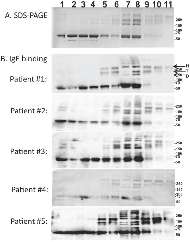
Panel A is an SDS-PAGE of rAra h 1: 1) untreated or heated in the presence of (lanes 2-6) glucose or (lanes 7-11) xylose for 1, 2, 4, 7 and 10 days, respectively. Panel B is an IgE Western blot analysis of 5 peanut allergic patient sera binding to the samples aligned with and described in panel A. Indicated by arrows are the location of the Ara h 1 hexamer (H), trimer (T) and dimer(D). Immunocap values for patients 1-5 are, respectively: >100, 23.1, 14.5, 21.5, and 37.1