
Figure 5. Identification of AGE-modified residues identified on the surface of Ara h 1.
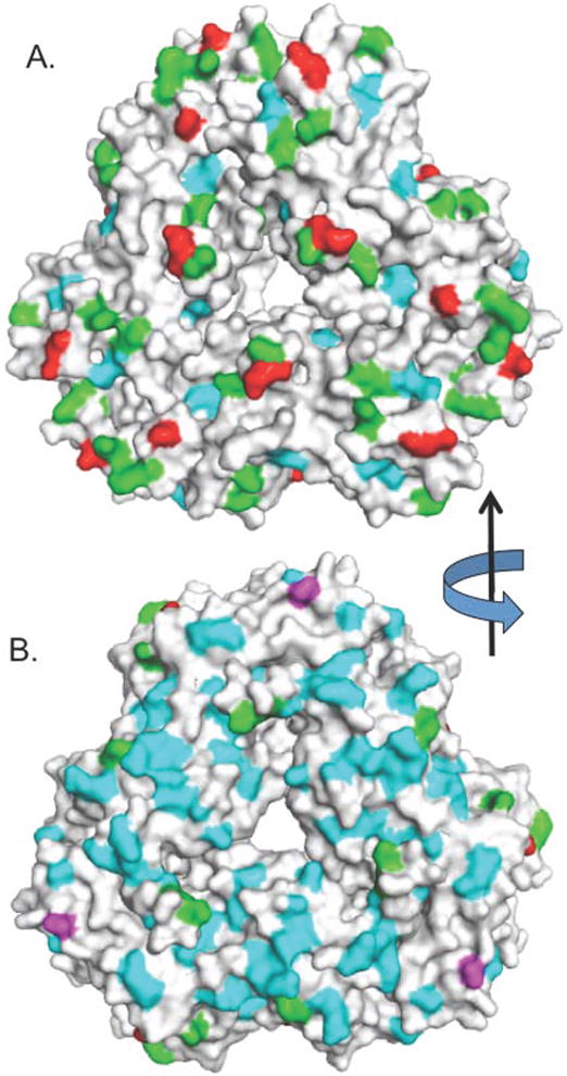
A surface rendering of Ara h 1 showing two views of the protein, rotated 180° about the y axis. Panel A is the “front” and panel B is the “back”. Unmodified lysines are colored green, modified lysines are red, umodified arginines are cyan, and modified arginines are magenta. The data are a composite of Tables 1 and 2.