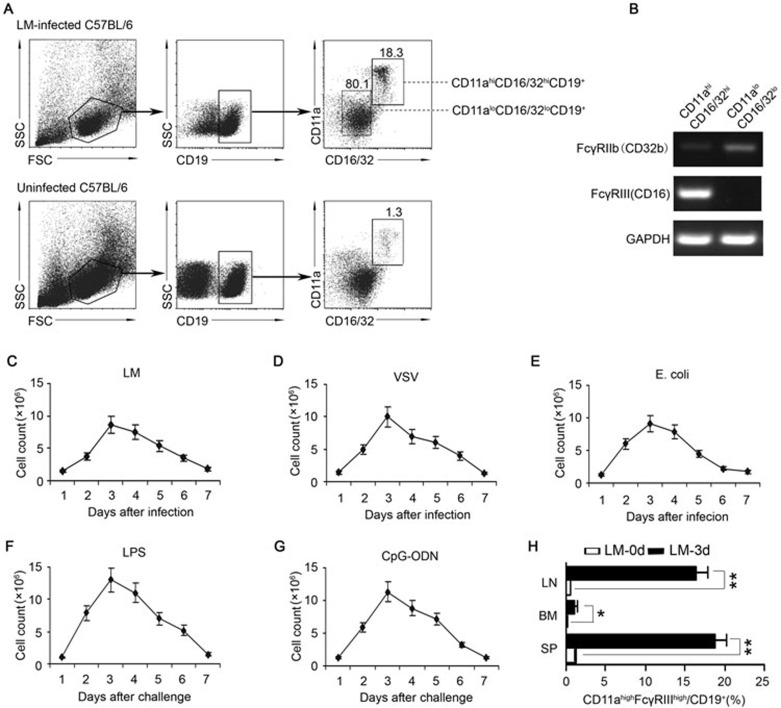
Figure 1.
Generation of CD11ahiFcγRIIIhiCD19+ cells in mice infected with pathogens or challenged with TLR ligands. Naïve C57BL/6 mice were i.p. infected with 2 × 10⁶ LM (A-C, H), 5 × 10⁶ PFU VSV (D), or 1 × 10⁶ E. coli (E). (A) Splenocytes were isolated on day 3 post-infection, and the percentage of CD11ahiFcγRIIIhiCD19+ cells in the CD19+ B cells was analyzed. (B) The FcγRIIb and FcγRIII mRNA expression of splenic CD11ahiFcγRIIIhiCD19+ or CD11aloFcγRIIIloCD19+ cells was assessed by RT-PCR. The transcript of the mouse GAPDH gene was used as an amplification control. (C-E) The number of CD11ahiFcγRIIIhi B cells in 108 splenocytes was examined within 7 days after infection with LM (C), VSV (D), and E. coli (E). (F, G) Naïve C57BL/6 mice were i.p. injected with LPS (0.5 mg/kg weight) (F) or CpG-ODN (2.5 mg/kg weight) (G). The numbers of CD11ahiFcγRIIIhi B cells in 108 splenocytes were dynamically examined within 7 days after the challenge. (H) The percentages of CD11ahiFcγRIIIhi B cells in the CD19+ B cells in the lymph nodes (LN), spleens (SP), and BM were analyzed on day 0 or day 3 post-LM infection. Data shown represent the mean ± SD. **P< 0.01, *P< 0.05.

