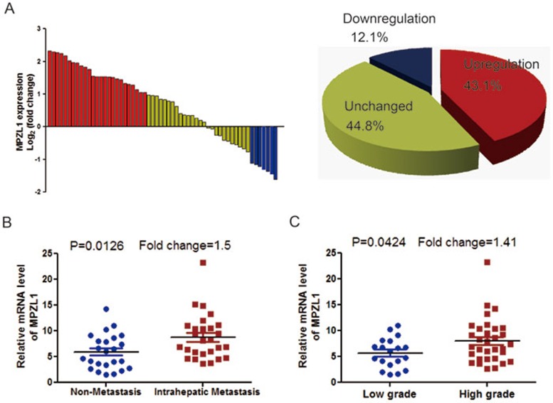
Figure 2.
Overexpression of MPZL1 correlates with malignant features of HCC. (A) The expression levels of MPZL1 in 58 paired HCC and matched non-tumor tissues were determined by q-PCR. The data are expressed as the log2 fold change (ΔCt [HCC/Non.]). Significant upregulation of MPZL1 expression in paired HCC/non-tumor samples was defined as a log2 fold change > 1 (i.e., 2-fold). The pie chart shows the proportions of HCC samples showing upregulation (red), downregulation (blue), and no change (yellow). (B) Overexpression of MPZL1 in primary HCCs with intrahepatic metastasis compared with that without intrahepatic metastasis according to the results of q-PCR. (C) Overexpression of MPZL1 in primary HCCs in high grade (grade = 3 and 4) compared with that in low grade (grade = 1 and 2) according to the results of q-PCR. Statistical analysis of differences between the two groups was performed by unpaired Student's t-test and P < 0.05 was considered statistically significant.

