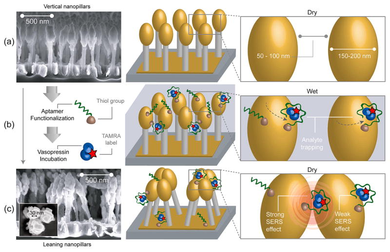
Figure 1.
Schematics of the functionalized leaning nanopillar detection method. (a) Highly packed gold-coated nanopillars are structured on a silicon wafer. Before exposure to the analyte, pillars are vertically oriented and are spaced by 50–100 nm gaps. (b) After aptamers are immobilized on the nanopillars (via Au–thiol covalent bonding) and a TVP sample is introduced, the analyte binds to the aptamers. (c) Scanning electron microscope (SEM) image of leaning nanopillars after the evaporation of liquid (side view). The analyte trapped within the leaning region exhibits a strong Raman enhancement effect due to the highly localized plasmonic field around the contact region. When the vasopressin is trapped by an aptamer within a hotspot, as shown on the right panel, the TAMRA Raman signal is highly enhanced. The inset shows a high-magnification SEM image displaying one of the pillar clusters, revealing junction spaces less than 10 nm between pillar heads (top view).

