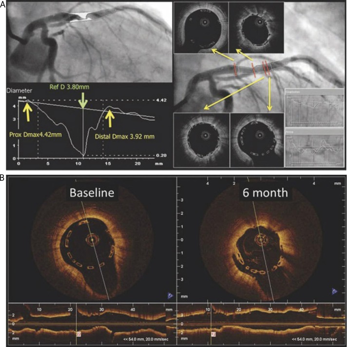
Fig. 5.
An example from the ABSORB cohort B: implantation of the 3.0 mm diameter BVS into the vessel with RVD diameter of 3.8mm (D max values in the proximal and distal references – 4.42 and 3.92mm respectively) (A). Undersizing of the vessel diameter leads to spontaneous malapposition at both ends of the BVS (B) at baseline (left) and after 6 months on OCT (with permission of Dr Gomez-Lara J) [17]

