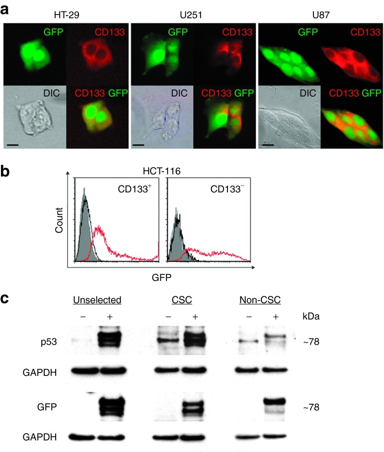
Figure 2.
scL-mediated exogenous gene expression in cancer stem-like cells (CSCs) in vitro. (a) Fluorescent and differential interference contrast (DIC) images of HT-29, U251, and U87 cells in monolayer culture transfected with scL-GFP in vitro for 48 hours. GFP-expressing cells were stained with an anti-CD133 antibody (red fluorescence). Scale bars indicate 10 μm. (b) Flow cytometry analysis of HCT-116 cells 48 hours after transfection with free GFP plasmid DNA (black open histograms) or scL-GFP (red open histograms). The cells were stained with CD133-PE antibody. GFP positivity in CD133+- and CD133−-gated cells are shown. Gray histograms represent untreated control. (c) Western blot analysis of HT-29 cells 24 hours after transfection with scL-GFP-p53. Expression of GFP and p53 fusion proteins in unselected, FACS-sorted CSCs (CD133+CD166+CD44+) and non-CSCs was assessed on two individual membranes. (−), untransfected cells; (+), scL-GFP-p53 transfected cells. FACS, flow-activated cell sorting.

