
Fig. 6.
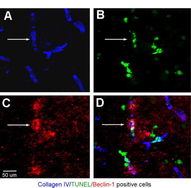
Ischemia-induced autophagic microvessel deterioration. (A–D) Immunohistochemical triple staining of collagen IV, Beclin-1 and TUNEL in the penumbra region 12 h after ischemia in a brain section from a p50−/− mouse. Collagen IV (blue) staining show consistent morphology of microvessels. Some of collagen IV-positive cells were co-labeled with TUNEL (green) and Beclin-1 (red) (arrows). The triple-positive staining indicated autophagic vasculature damage. (For interpretation of the references to color in this figure legend, the reader is referred to the web version of this article.)