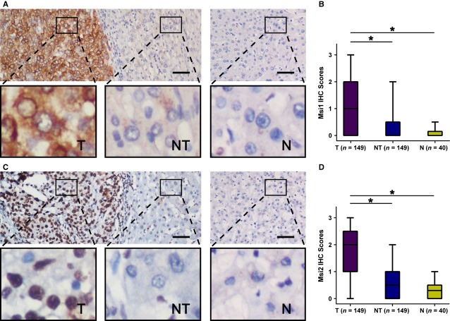
Figure 1.
Musashi1 (MSI1) and MSI2 were significantly up-regulated in hepatocellular carcinoma (HCC). (A) Immunohistochemistry (IHC) assays of MSI1 expression in 40 normal hepatic tissues and 149 paired HCC and adjacent non-tumourous tissues. The upper left panel represents paired HCC and non-tumourous tissues and reveals high MSI1 expression in HCC tissues and low expression in adjacent non-tumourous tissues. The upper right panel represents normal hepatic tissues and reveals low MSI1 expression. Scale bars, 50 μm. Lower panels represent magnified pictures of boxed area in the corresponding upper panels. (B) MSI1 expression levels were compared among HCC, adjacent non-tumourous and normal hepatic tissue specimens. *P < 0.001. (C) IHC assays of MSI2 expression in 40 normal hepatic tissues and 149 paired HCC and adjacent non-tumourous tissues. The upper left panel represents paired HCC and non-tumourous tissues and was interpreted as high MSI2 expression in HCC tissues and low expression in adjacent non-tumourous tissues. The upper right panel represented normal hepatic tissues and was interpreted as low expression of MSI2. Scale bars, 50 μm. Lower panels represent magnified pictures of boxed area in the corresponding upper panels. (D) MSI2 expression levels were compared among HCC, adjacent non-tumourous and normal hepatic tissues. *P < 0.001.

