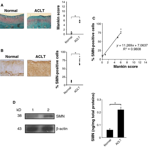
Figure 5.

Survival of motor neuron (SMN) expression levels in an experimental osteoarthritis (OA) model in vivo. (A) Safranin O staining of histological sections from rabbit knee joint cartilage (representative samples; view of the femoral condyle; magnification ×4) and grading of OA severity (all samples were processed for the analyses). (B) SMN-specific immunoreactivity on sections from rabbit knee joint cartilage (representative samples; view of the femoral condyle; magnification ×20) and percentages of SMN-positive cells (all samples were processed for the analyses) (normal cartilage: 7.1–13.7%; OA cartilage: 59.7–83.8% as for moderate human OA). (C) Correlation between the SMN expression levels and OA severity (combination of all values from A and B). (D) Analysis of SMN by Western blotting (lane 1, normal cartilage; lane 2, ACLT cartilage; all representative samples) and ELISA (SMN concentrations in all freshly isolated normal and ACLT chondrocytes; *statistically significant difference).
