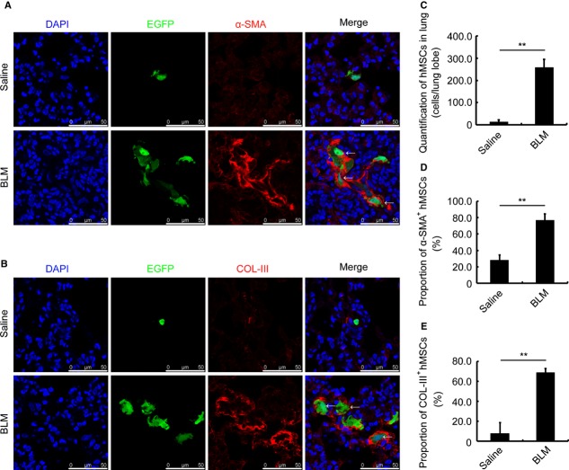
Figure 6.
hBMSCs are involved in lung injury of SCID/Beige mice. (A) Immunofluorescence staining for α-SMA (red) in the lung of Saline- (upper) or BLM-(lower) challenged SCID/Beige mice. The mice were transplanted with GFP-labelled hBMSCs 48 hrs after bleomycin administration. DAPI (blue) staining shows the nucleus. α-SMA-positive cells are indicated with arrows. (B) Immunofluorescence staining for collagen III (COL-III, red) in the lung of Saline- (upper) or BLM-(lower) challenged SCID/Beige mice. The mice were transplanted with GFP-labelled hBMSCs 48 hrs after bleomycin administration. DAPI (blue) staining shows the nucleus. The COL-III-positive cells are indicated with arrows. (C) Quantification of accumulated hBMSCs in the lung of Saline- and BLM-treated mice. (D) Proportion of α-SMA-positive hBMSCs in the lung of Saline- and BLM-treated mice. (E) Proportion of COL-III-positive hBMSCs in the lung of Saline- and BLM-treated mice. All the lung tissues were isolated at day 7 after saline or BLM administration. Each value represents the mean ± SE. n = 6 mice in each group. **P < 0.01. BLM: bleomycin.

