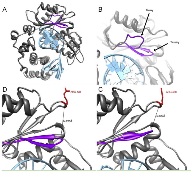
Fig. 3.
(A) Overlay of Polλ in the binary and ternary conformations. DNA is shown in light blue, and the Loop 1 is shown in purple. (B) Differences in Loop 1 orientation between the two conformations. Distance between position 438 and Loop 1 following an interpolation between the two structures at its furthest (Panel D) and closest (Panel C) approaches. (For interpretation of the references to color in this artwork, the reader is referred to the web version of the article.)

