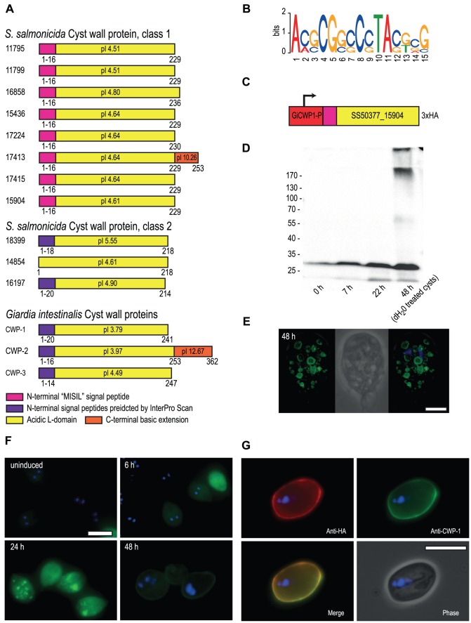
Figure 2. S. salmonicida cyst wall proteins traffic in G. intestinalis ESVs and incorporate into the cyst wall.
A. Schematic representation of candidate S. salmonicida cyst wall proteins (CWPs). Numbers refer to protein IDs and conserved features are shown by coloured boxes with the amino acid positions indicated. The isoelectric point (pI) is indicated. B. Sequence logo of motifs upstream of the eight class 1 CWPs, glucosamine-6 phosphate deaminase, two glucose 6-phosphate N-acetyltransferase and two UDP-glucose 4-epimerases. C. The construct used to express the candidate S. salmonicida class 1 CWP in G. intestinalis during encystation. The red box indicates the promoter region of G. intestinalis CWP-1. D. Western blot of samples taken from G. intestinalis transfectants carrying the S. salmonicida CWP construct. Expected size of the epitope-tagged SS50377_15904 is 28.4 kDa. E and F. Immunofluorescence analysis of G. intestinalis transfectants at different time points into encystation. The protein was detected using anti-HA conjugated to AlexaFluor488 (green) and the nuclei were labelled using DAPI (blue). Scale bars, 5 µm. G. Immunofluorescence micrograph of a G. intestinalis transfectant at 48 h post inducation of encystation following water-treatment. The cysts were stained by rabbit anti-HA and detected by anti-rabbit Alexa Fluor 594 (red), and probed using anti-CWP-1 conjugated to FITC (green). Scale bar, 10 µm.

