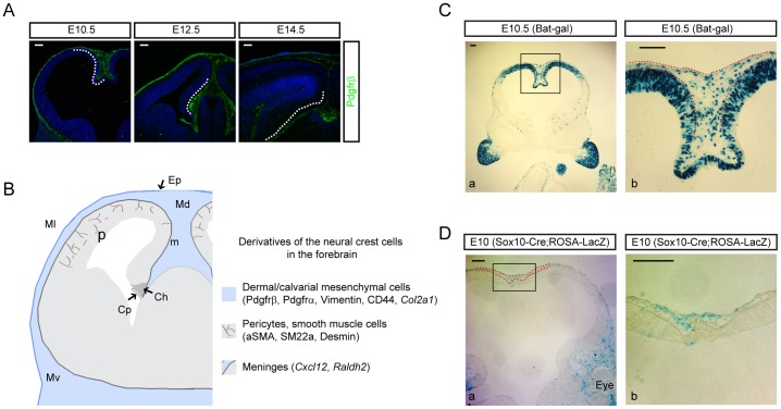
Figure 1. Wnt-responding mesenchymal cells expand in the dorsal interhemispheric region.
A) Staining of Pdgfrß+ neural crest-derived mesenchymal cells from E10 to E14. Dashed lines highlight the dorsomedial mesenchymal cells, which expand at E10 and spread laterally at later ages. At E14, perivascular cells strongly express Pdgfrß. B) A diagram showing mesenchymal cells at the level of dorsoventral axis in the forebrain; the dorsal (Md), lateral (Ml), and ventral (Mv) mesenchymal cells. The derivatives of the neural crest cells are listed with markers used in this study. Ep = epidermis, Ch = cortical hem, Cp = choroid plexus, P = pericytes, m = meninges. C) X-gal staining of E10.5 Bat-gal transgenic embryos. A high power image of the boxed area of C-a is presented in C-b. X-gal+ mesenchymal cells are localized in the interhemispheric region (red dashed lines). D) X-gal staining of E10 ROSA-lacZ Cre reporter mice crossed with Sox10-Cre, a neural crest driver. A high magnification image of the boxed area in D-a is shown in D-b. Red dashed lines mark the area with mesenchymal cells. Scale bars = 100 µm.

