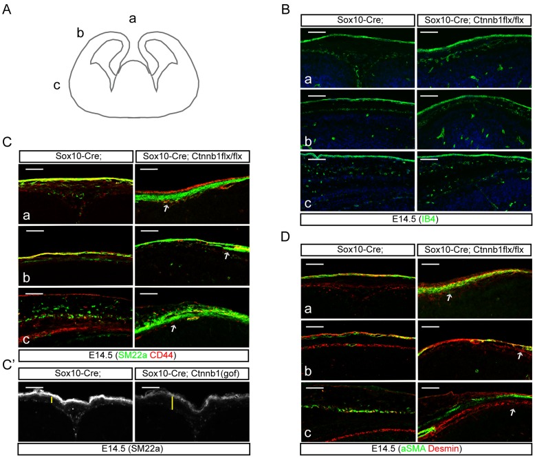
Figure 7. Ectopic generation of smooth muscle cells by loss of β-catenin in neural crest cells.
A) A schematic drawing shows the three regions (a, b, and c) used to stain markers for smooth muscle cells. B) Isolectin IB4 staining shows distribution of blood vessels in the epidermis and meninges. Fewer mesenchymal cells were seen in the space between the blood vessels of Sox10-Cre;Ctnnb1(lof)flx/flx mutant embryos. C) SM22a, a marker for the smooth muscle cells, and CD44, a marker for MSCs, were used to characterize the mesenchymal cells in the regions a–c. C′) Sox10-Cre;Ctnnb1(gof) mutant embryos double-stained for SM22a and CD44. Yellow bars indicate the thickness of the mesenchyme. D) aSMA, a marker for smooth muscle cells, and Desmin, a marker for pericytes, were used to reveal the ectopic generation of smooth muscle cells from neural crest cells of Sox10-Cre;Ctnnb1(lof)flx/flx mutant embryos at E14.5. Arrows indicate the incompetent spreading of mesenchymal cells into the ‘b’ region of Sox10-Cre;Ctnnb1(lof)flx/flx mutants. Scale bars = 100 µm.

