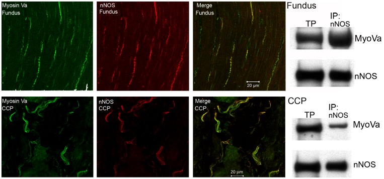
Figure 1. Localization and interaction of myosin Va and nNOS in the fundus and corpora cavernosa (CCP).
In both fundus (top panels) and CCP (bottom panels), immunoreactivity for myosin Va (green) is present in nerve fibers that are immunoreactive for nNOS (red). Co-localization (yellow) is present in projected image. Western blot on right shows positive bands for myosin Va and nNOS in total protein lysate (TP) in lane 1. In lane 2, samples were immunoprecipitated for nNOS and blotted for myosin Va to demonstrate molecular interaction between these proteins in fundus (top panel) and CCP (bottom panel).

