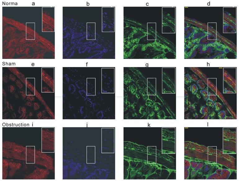
Figure 8. Immunofluorescence staining of KV2.2 in the mice small intestine frozen section.
The cell membrane was stained by Fm 4-64 membrane stain (red fluorescence) and cell nuclear was stained by DAPI (blue fluorescence). The green fluorescence was KV2.2 immunofluorescence. Scale bar = 20 µm, the experiments repeated six times.

