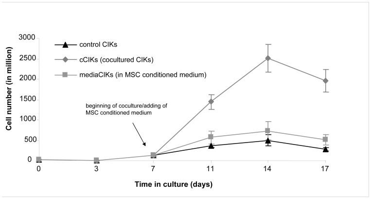
Figure 1. Comparison of cytokine-induced killer cell (CIK) proliferation.
CIKs were cultivated according to protocol (controls). On day 7, some of the CIKs were cultivated either with mesenchymal stem cells (cCIKs) or in MSC-conditioned medium (mediaCIKs), and proliferation was compared with that of controls. After starting the coculture, cCIKs proliferated faster than controls (p<0.05). The mediaCIKs displayed increased proliferation (p>0.1), too, but proliferation rates did not reach the level of cCIK proliferation.

